
Central Venous & Iliocaval Ultrasound in New Jersey
Universal Ultrasound Diagnostics provides advanced central venous and iliocaval ultrasound in Flemington, New Jersey. This specialized examination evaluates major veins in the abdomen and pelvis — including the inferior vena cava (IVC) and iliac veins — to assess blood flow and look for findings such as thrombosis, obstruction, or venous compression.
Difficulty breathing, chest pain or discomfort, coughing up blood, a fast or irregular heartbeat, severe lightheadedness, or fainting - these can be signs of pulmonary embolism. Call 911 or go to the nearest emergency department immediately.
What is central venous ultrasound?
Central venous ultrasound extends the venous examination beyond the legs to evaluate the large veins that carry blood from the pelvis and lower body back toward the heart.
What does UUDVT evaluate?
Why might this test be ordered?
Central venous imaging may be useful when symptoms, prior DVT history, abnormal lower-extremity Doppler findings, swelling patterns, or a clinician's concern suggest disease above the groin.
What happens during the exam?
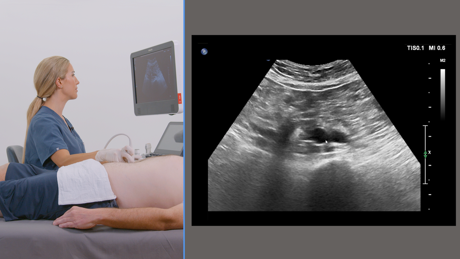
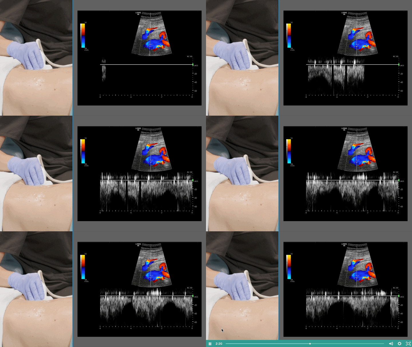
A credentialed technologist places ultrasound gel on the abdomen and pelvis and uses a transducer to evaluate the IVC and iliac veins. Because these vessels sit deeper than the leg veins, imaging relies more heavily on Doppler assessment of flow, and you may be asked to breathe, hold your breath, or bear down briefly to help evaluate venous flow patterns.
Gel & positioning
Ultrasound gel is applied and you're positioned comfortably for imaging of the abdomen and pelvis.
IVC & iliac vein imaging
The technologist images the inferior vena cava and iliac venous segments, following our accredited protocol.
Doppler flow assessment
Doppler ultrasound evaluates venous flow patterns and looks for findings suggesting obstruction or compression.
Areas examined, timing & results
What areas are examined?
Imaging covers the inferior vena cava and iliac venous segments, following our accredited protocol and the exact study ordered by your clinician.
How long does the test take?
Usual test lasts 45 minutes. Body habitus, bowel gas, anatomy, and exam complexity can affect imaging time.
What happens after the ultrasound?
Reports are sent directly to your doctor quickly and securely.
Stat results are provided immediately, routine results within 24 hours.
Frequently Asked Questions
What is an iliocaval ultrasound?
It is an ultrasound examination of the iliac veins in the pelvis and the inferior vena cava in the abdomen.
Why would my doctor look above the leg veins?
Some venous problems are located in the pelvis or abdomen. Evaluating central veins may provide additional information when lower-extremity symptoms or Doppler findings suggest a more proximal issue.
Can ultrasound evaluate iliac vein compression?
Ultrasound can assess anatomy and venous flow for findings that may suggest compression or obstruction. Whether additional imaging is needed is determined by the treating clinician.
Is this the same as a standard DVT ultrasound?
No. A standard lower-extremity DVT study focuses primarily on the leg veins. Central venous/iliocaval imaging evaluates veins in the pelvis and abdomen.
Do I need an order?
Policy varies by insurance-billed versus self-pay testing: you don't need a physician order if you're self-pay testing, you'll need an order if you go through insurance, with appropriate CPT code and symptomatology ICD 10 code
Need Central Venous Imaging?
Schedule online or call/text our Flemington office. If you have emergency symptoms, call 911.